Cerebrovascular Disease
Anatomy of the Cerebral Circulation
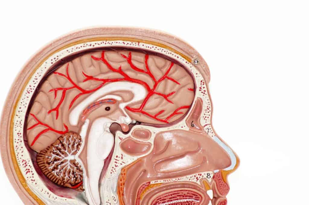
In dogs, the brain receives its blood supply primarily through the basilar artery and the internal carotid arteries. These vessels unite at the base of the brain to form the arterial circle, commonly known as the Circle of Willis. This vascular network plays an essential role in maintaining continuous cerebral perfusion and protecting brain tissue from ischemic injury.
Three major pairs of cerebral arteries originate from the arterial circle and distribute blood throughout different brain regions:
- The rostral cerebral arteries supply the rostral and medial portions of the cerebrum.
- The middle cerebral arteries, which are the largest cerebral vessels, perfuse the lateral cerebral hemispheres as well as significant portions of the basal nuclei and thalamus.
- The caudal cerebral arteries provide blood to the caudal and caudomedial cerebral hemispheres and the rostral cerebellum.
Additional branches from the basilar artery vascularize the caudal cerebellum, pons, and medulla oblongata. Variations in the anatomy of the Circle of Willis are frequently observed in dogs and may influence susceptibility to cerebrovascular disorders, similar to what is documented in human medicine.
The cerebral circulation is protected by collateral vascular pathways that preserve blood flow during arterial obstruction. The arterial circle allows communication between the carotid and vertebrobasilar systems, while secondary extracranial collateral vessels can develop between the external carotid and vertebral arteries when carotid obstruction occurs. Small intracranial anastomoses are also present on the cortical surface; however, these vessels are often insufficient to compensate for occlusion of a major cerebral artery, potentially leading to ischemia and infarction.
In cats, cerebral vascular anatomy differs significantly from dogs. Shortly after birth, the proximal internal carotid artery regresses, and cerebral circulation becomes dependent on branches of the external carotid artery, particularly the maxillary and pharyngeal arteries. Blood flow within the basilar artery is directed away from the brain, and the vertebral arteries mainly supply the caudal brain stem.
Regulation of Cerebral Blood Flow
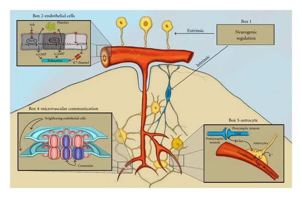
The brain possesses highly specialized autoregulatory mechanisms that maintain stable cerebral blood flow despite fluctuations in systemic circulation. Cerebral arterioles continuously adjust their diameter in response to physiologic and metabolic changes.
When systemic arterial pressure increases, cerebral arterioles constrict to prevent excessive perfusion and vascular injury. Conversely, reduced arterial pressure induces vasodilation to preserve oxygen and nutrient delivery to neural tissue.
Carbon dioxide is one of the most important regulators of cerebral circulation. Elevated arterial carbon dioxide tension (PaCO₂) causes cerebral vasodilation and increases cerebral blood flow, whereas reduced PaCO₂ induces vasoconstriction and decreases perfusion. Oxygen concentration also influences vascular tone, although significant vasodilation usually occurs only when arterial oxygen tension falls below approximately 50 mm Hg.
Regional cerebral blood flow is tightly coupled to metabolic activity. Brain regions with increased neuronal activity require greater oxygen and glucose delivery, resulting in localized vasodilation and increased perfusion. Conditions such as seizures markedly elevate cerebral metabolic demand and consequently produce significant increases in cerebral blood flow.
In animals affected by cerebrovascular disease, these autoregulatory and metabolic control mechanisms may become impaired, contributing to ischemia, edema, and progressive neurologic dysfunction.
Pathophysiology of Cerebral Ischemia and Infarction
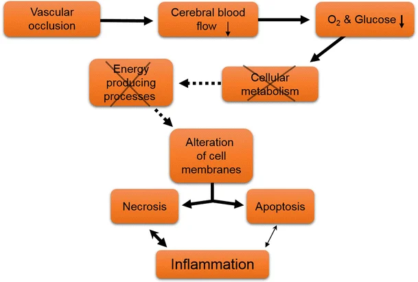
Cerebral ischemia occurs when blood flow decreases below the level required for normal neuronal function. If the reduction in perfusion becomes severe or prolonged, irreversible tissue necrosis develops, resulting in cerebral infarction.
The severity and duration of ischemia are the principal determinants of neuronal survival. Experimental studies in cats demonstrate that when cerebral blood flow falls from normal levels of approximately 35 mL/100 g/min to 15–18 mL/100 g/min, neuronal electrical activity ceases while cellular viability is temporarily preserved. When perfusion drops below 10–12 mL/100 g/min, disruption of ionic homeostasis occurs and irreversible injury rapidly develops.
Temporary vascular occlusion may produce reversible neurologic deficits if circulation is restored rapidly. Short-term occlusion of the middle cerebral artery may cause transient dysfunction with minimal infarction, whereas prolonged occlusion results in permanent neurologic deficits associated with extensive tissue necrosis.
Cellular Mechanisms of Ischemic Brain Injury
The primary mechanism of neuronal injury during cerebral infarction is failure of cellular energy metabolism. Reduced oxygen and glucose delivery impair synthesis of adenosine triphosphate (ATP), which is essential for maintaining ion gradients across neuronal membranes.
Loss of ATP initiates several destructive cellular processes:
- Failure of sodium-potassium pumps causes intracellular accumulation of sodium, chloride, and water, leading to cytotoxic edema.
- Anaerobic glycolysis increases lactic acid production and intracellular acidosis.
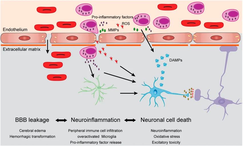
- Excess intracellular calcium activates proteases and phospholipases that degrade structural proteins and membrane phospholipids.
- Phospholipid degradation generates free radicals that damage proteins, lipids, and cellular membranes.
These combined events progressively disrupt neuronal integrity and ultimately lead to irreversible necrosis. Current research suggests that therapies targeting calcium influx, oxidative stress, and free radical formation may help reduce ischemic injury.
Arterial Thrombosis and Embolism
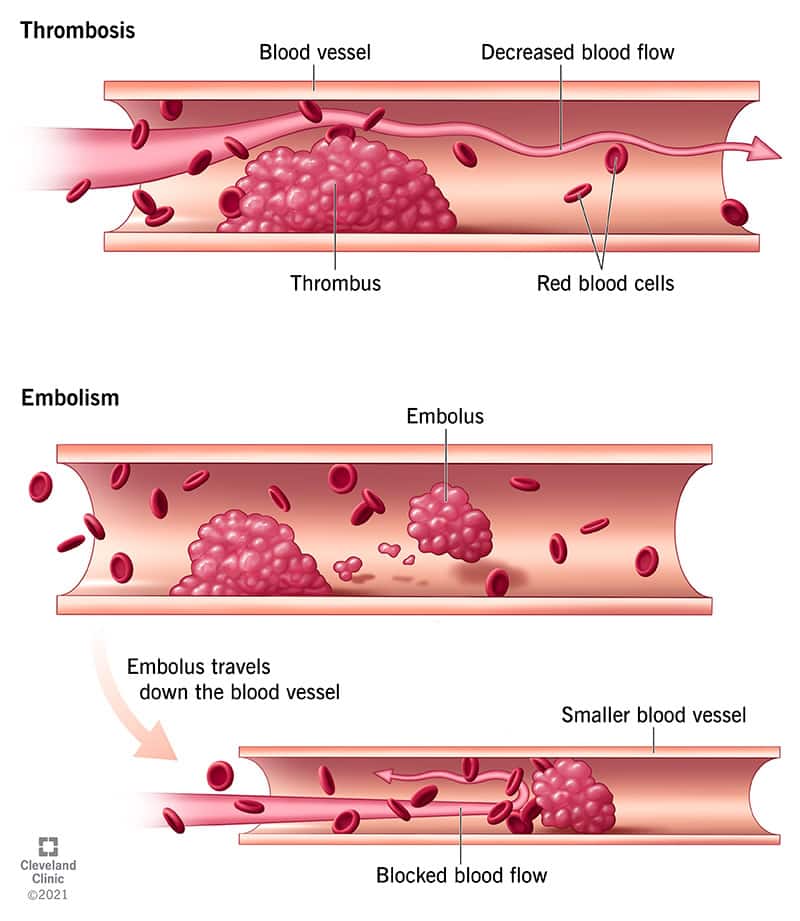
Focal cerebral ischemia commonly results from thrombosis or embolism.
- A thrombus is a blood clot that forms directly within a vessel and obstructs local blood flow.
- An embolus is material, usually a clot fragment, that travels through the bloodstream and lodges within a distant cerebral vessel.
In human medicine, atherosclerosis is the leading cause of thromboembolic stroke. Atherosclerotic degeneration damages the vascular endothelium, promotes platelet aggregation, and predisposes to thrombus formation and embolization.
Atherosclerosis also occurs in dogs, especially in:
- older animals,
- dogs with hypothyroidism,
- and Miniature Schnauzers affected by hyperlipoproteinemia.
Additional disorders associated with cerebral thromboembolism in dogs include:
- sepsis,
- coagulopathies,
- neoplasia,
- and heartworm disease.
Feline Ischemic Encephalopathy
Feline ischemic encephalopathy is a syndrome characterized by acute focal brain infarction in cats. Clinical signs commonly include:
- hemiparesis,
- blindness,
- behavioral abnormalities,
- and seizures.
Although thrombosis has occasionally been identified, vascular lesions are often absent. One proposed mechanism involves vasospasm induced by aberrant migration of parasitic larvae such as Cuterebra species.
Some affected cats recover partially over several weeks, while severe cases may be fatal.
Venous Infarction
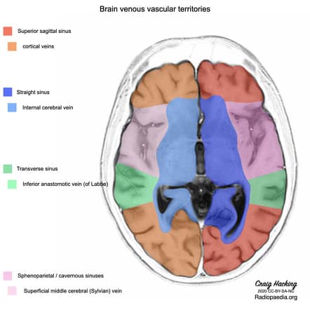
Venous infarction develops when cortical veins or dural venous sinuses become obstructed by thrombosis, inflammation, neoplasia, or trauma.
Because venous collateral circulation is extensive, venous infarction is less common than arterial infarction. However, preserved arterial inflow combined with impaired venous drainage often results in severe edema and hemorrhage.
Spontaneous Intracranial Hemorrhage
Spontaneous intracranial hemorrhage refers to nontraumatic bleeding within or around the brain. Hemorrhage may occur in several locations:
- epidural,
- subdural,
- subarachnoid,
- intraparenchymal,
- or intraventricular spaces.
Primary intraparenchymal hemorrhage is frequently associated with systemic hypertension and vascular degeneration. Secondary hemorrhage may occur due to:
- cerebral infarction,
- vascular malformations,
- amyloid angiopathy,
- neoplasia,
- vasculitis,
- or coagulation disorders.
Hemorrhagic infarction often develops when ischemic tissue undergoes reperfusion through collateral circulation or spontaneous thrombolysis, allowing blood leakage through damaged vessels.
Cerebral Vascular Malformations
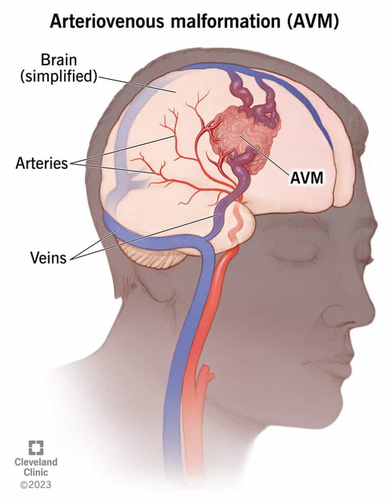
Congenital vascular malformations can predispose animals to intracranial hemorrhage. Major categories include:
- arteriovenous malformations,
- venous malformations,
- capillary telangiectasia,
- and cavernous malformations.
Arteriovenous malformations are particularly important because direct arterial-to-venous shunting exposes fragile vessels to high pressure and increases the risk of hemorrhage.
Clinical signs often do not appear until middle age or later, when vascular rupture occurs.
Clinical Signs of Cerebrovascular Disease
The hallmark of cerebrovascular disease is the sudden onset of focal neurologic dysfunction. Clinical manifestations depend on lesion location and severity.
Common signs include:
- seizures,
- hemiparesis,
- circling,
- blindness,
- cranial nerve deficits,
- ataxia,
- vestibular dysfunction,
- altered consciousness,
- and coma.
Forebrain lesions often produce contralateral postural deficits and behavioral abnormalities, whereas cerebellar lesions may cause hypermetria and vestibular signs. Brain stem involvement is associated with severe neurologic dysfunction including tetraplegia and impaired consciousness.
Neurologic deterioration during the first few days may result from progressive cerebral edema and increased intracranial pressure.
Diagnostic Evaluation
Diagnosis of cerebrovascular disease requires a combination of neurologic examination, laboratory testing, and advanced neuroimaging.
Important diagnostic procedures include:
- complete blood count,
- serum biochemistry,
- coagulation testing,
- blood pressure measurement,
- cerebrospinal fluid analysis,
- electrocardiography,
- thoracic radiography,
- CT imaging,
- and MRI.
Computed tomography is highly effective for detecting acute intracranial hemorrhage, while magnetic resonance imaging is more sensitive for early ischemic infarction and brain stem lesions.
Advanced imaging also helps differentiate vascular disease from neoplasia, encephalitis, trauma, and other focal brain disorders.
Treatment of Cerebrovascular Disease
Therapeutic management focuses on preserving cerebral oxygenation, reducing secondary injury, and treating underlying disease.
Maintenance of Cerebral Perfusion
Adequate ventilation and oxygen delivery are essential. Animals with impaired consciousness may require endotracheal intubation and mechanical ventilation.
Hypotension and dehydration should be corrected carefully because reduced cerebral perfusion pressure worsens ischemia. Excessive crystalloid administration should be avoided since it may aggravate cerebral edema.
Control of Intracranial Pressure
Elevated intracranial pressure can compromise cerebral perfusion and induce brain herniation.
Common therapeutic measures include:
- controlled hyperventilation,
- mannitol administration,
- osmotic therapy,
- and head elevation.
Mannitol improves cerebral perfusion by reducing blood viscosity and promoting osmotic movement of water out of brain tissue.
Hyperventilation lowers PaCO₂, causing cerebral vasoconstriction and temporary reduction of intracranial blood volume.
Seizure Management
Seizures substantially increase cerebral metabolic demand and intracranial pressure. Rapid seizure control is therefore critical.
Common anticonvulsant therapies include:
- diazepam for emergency seizure control,
- and phenobarbital for maintenance therapy.
Prognosis
Prognosis depends primarily on:
- lesion size,
- lesion location,
- severity of edema,
- intracranial pressure,
- and the presence of underlying systemic disease.
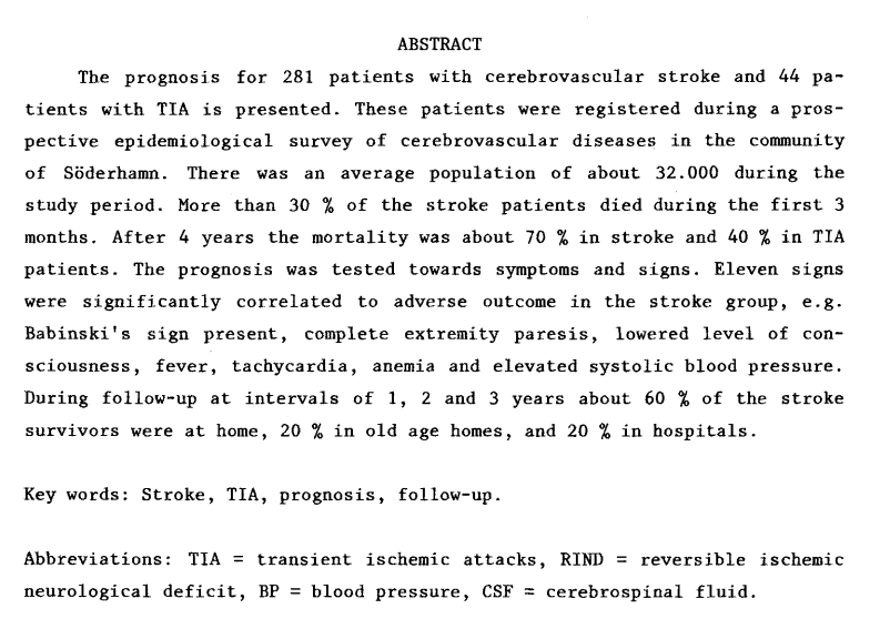
Animals with mild cerebral infarction may recover significantly over several weeks, whereas severe hemorrhage, uncontrolled seizures, brain herniation, or persistent coma carry a guarded prognosis.
With rapid diagnosis, supportive care, and treatment of underlying disorders, many animals affected by cerebrovascular disease can experience meaningful neurologic recovery.
