Regulation of Cerebral Circulation
Introduction to Cerebral Blood Flow Regulation
The regulation of cerebral circulation is a highly complex physiological process that ensures continuous oxygen and nutrient delivery to brain tissue. Because the brain has a high metabolic demand and limited energy reserves, cerebral blood flow (CBF) must remain tightly controlled under both normal and pathological conditions. Multiple regulatory systems cooperate to maintain vascular homeostasis, including neurogenic regulation, chemical regulation, metabolic control, and autoregulatory mechanisms. These systems collectively preserve cerebral perfusion, stabilize intracranial microcirculation, and protect neural tissue from ischemic or hypertensive injury.
Neurogenic Regulation of Cerebral Circulation
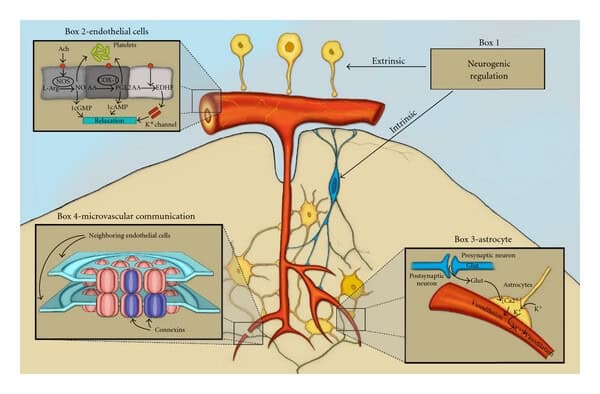
Neurogenic regulation of cerebral blood flow has been extensively investigated for several decades. Although important discoveries have clarified many aspects of cerebrovascular innervation, the exact physiological contribution of vasomotor nerves to cerebral perfusion control remains incompletely understood.
Sympathetic Adrenergic Innervation
Cerebral blood vessels receive adrenergic innervation primarily from fibers originating in the superior cervical ganglion. These sympathetic fibers supply both pial arteries and intracerebral vessels. However, compared with peripheral vascular beds, cerebral vessels exhibit relatively weak responses to sympathetic stimulation.
Experimental studies in cats demonstrated that electrical stimulation of cervical sympathetic nerves induces modest vasoconstriction of pial arterioles, generally ranging between 7% and 12%. Larger cerebral arteries show stronger constrictive responses than smaller arterioles. In many cases, vessels smaller than 100 µm display minimal or no measurable response to adrenergic stimulation.
The magnitude of sympathetic cerebrovascular responses varies significantly among species. Monkeys and rabbits exhibit the most consistent reductions in cerebral blood flow during sympathetic activation, whereas cats and dogs display weaker or inconsistent effects. In rabbits, vasoconstriction is often transient because of vascular escape mechanisms that restore blood flow despite ongoing sympathetic stimulation.
The relatively limited responsiveness of cerebral vessels to sympathetic stimulation is mainly attributed to the low sensitivity of cerebrovascular alpha-adrenergic receptors to norepinephrine. Developmental and embryological characteristics of cerebral vessels may also contribute to this reduced adrenergic responsiveness.
Importantly, cerebral sympathetic nerves do not appear to exert significant resting vasomotor tone under normal physiological conditions.
Sympathetic Protection During Hypertension
Although sympathetic effects on cerebral circulation are usually modest, their importance increases substantially during severe arterial hypertension. When mean arterial pressure rises to approximately 200 mm Hg, stimulation of sympathetic nerves produces more pronounced vasoconstriction in cats, dogs, and monkeys.
This enhanced vasoconstrictor response may serve a protective function by limiting excessive pressure transmission to delicate cerebral capillaries and small arterioles. Such protection helps preserve blood-brain barrier integrity and reduces the risk of vascular damage or cerebral edema during hypertensive episodes.
Small cerebral arterioles possess thin vascular walls and relatively limited smooth muscle content, making them particularly vulnerable to pressure-induced injury. Sympathetic constriction of larger upstream vessels may therefore reduce downstream microvascular stress.
Reflex Neurogenic Control
The participation of sympathetic nerves in reflex regulation of cerebral blood flow remains controversial. Most experimental evidence suggests that reflex neurogenic influences play only a limited role in resting cerebrovascular regulation.
Studies involving carotid baroreceptor stimulation, chemoreceptor activation, or denervation procedures generally show little effect on resting cerebral blood flow, cerebrovascular resistance, or vascular responsiveness to carbon dioxide and hypoxia.
Some evidence nevertheless indicates minor reflex contributions. For example:
- Carbon dioxide inhalation can activate sympathetic nerves and partially oppose CO₂-induced vasodilation in larger pial arteries.
- Severe hypotension associated with hemorrhage may trigger sympathetic vasoconstriction that further reduces cerebral blood flow.
- Hypercapnia-induced vasodilation may become greater after cervical sympathectomy, suggesting that sympathetic activity normally exerts mild counter-regulatory vasoconstriction.
Overall, available evidence indicates that reflex sympathetic mechanisms have only secondary importance in physiological regulation of cerebral circulation.
Cholinergic Regulation of Cerebral Vessels
Unlike intracerebral vessels, pial arteries receive cholinergic innervation. Activation of these cholinergic fibers produces cerebral vasodilation through stimulation of vascular cholinergic receptors.
Electrical stimulation of cranial nerves such as the facial nerve and greater superficial petrosal nerve can modestly increase cerebral blood flow, likely through activation of cholinergic vasodilator pathways.
However, the physiological relevance of these cholinergic mechanisms remains uncertain. Experimental denervation studies demonstrate that removal of cholinergic input does not significantly impair cerebral autoregulation or vascular responses to hypoxia and hypercapnia.
Consequently, cholinergic pathways are not considered major regulators of resting cerebral circulation.
Brainstem Vasomotor Control
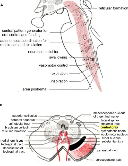
Several studies proposed that vasomotor centers within the brainstem, particularly the locus coeruleus, may regulate cerebral blood flow through direct neural control of cerebral vessels.
Experimental stimulation or ablation of brainstem regions has produced alterations in cerebral blood flow, autoregulatory responses, and vascular permeability. However, these findings remain controversial because many observed effects may result indirectly from changes in systemic blood pressure or neuronal metabolic activity rather than direct vasomotor innervation.
Current evidence does not conclusively support the existence of dominant brainstem-mediated neurogenic control of cerebral circulation.
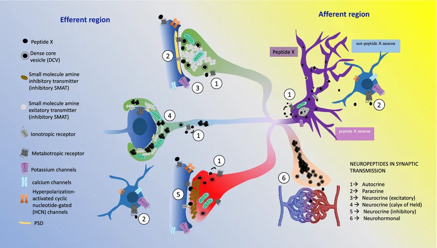
Peptidergic Innervation and VIP
Peptidergic nerve fibers containing vasoactive intestinal polypeptide (VIP) have been identified in pial arteries. VIP is a potent vasodilator capable of relaxing cerebral vascular smooth muscle.
Although these fibers may contribute to cerebrovascular regulation, their exact physiological role remains poorly understood and requires further investigation.
Chemical Regulation of Cerebral Blood Flow
Carbon Dioxide and pH Regulation
Carbon dioxide is one of the most powerful physiological regulators of cerebral circulation. Increases in arterial CO₂ concentration (hypercapnia) produce marked cerebral vasodilation, increased cerebral blood flow, and reduced cerebrovascular resistance. Conversely, hypocapnia induces vasoconstriction and decreases cerebral perfusion.
Smaller cerebral arterioles are particularly sensitive to hypercapnia and exhibit greater vasodilatory responses than larger vessels.
Mechanism of CO₂ Action
The primary mechanism underlying CO₂-induced vasodilation involves direct effects on vascular smooth muscle mediated by extracellular hydrogen ion concentration (pH). Molecular CO₂ itself and bicarbonate ions possess little intrinsic vasoactivity.
Changes in pH surrounding cerebral vessels strongly influence vascular caliber. Because CO₂ readily crosses the blood-brain barrier while bicarbonate diffusion is limited, alterations in arterial CO₂ significantly modify cerebrospinal fluid pH and thereby regulate vascular tone.
Local tissue acidosis during hypercapnia relaxes vascular smooth muscle and promotes vasodilation.
Adaptation to Chronic Hypercapnia and Hypoxia
During prolonged exposure to abnormal respiratory conditions, cerebrospinal fluid bicarbonate concentration gradually changes to compensate for pH disturbances.
- Chronic hypercapnia increases bicarbonate levels and reduces cerebrovascular sensitivity to CO₂.
- Chronic hypoxia decreases bicarbonate concentration and enhances vasodilatory responsiveness to hypercapnia.
These adaptive responses are also observed during high-altitude exposure and contribute to long-term cerebral blood flow regulation.
Hypoxia-Induced Vasodilation
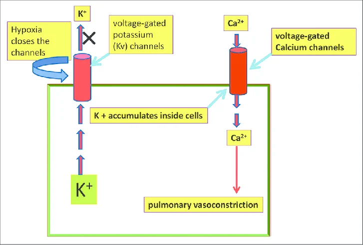
Arterial hypoxia produces cerebral vasodilation, increased cerebral blood flow, and decreased vascular resistance. Similar effects occur during carbon monoxide exposure.
Experimental evidence indicates that hypoxic vasodilation is mediated primarily by local tissue mechanisms rather than neural pathways. Reduced oxygen availability directly alters vascular smooth muscle behavior and stimulates production of vasodilatory metabolites.
Metabolic Regulation of Cerebral Blood Flow
The brain depends heavily on aerobic metabolism and therefore requires precise coupling between neuronal activity and regional blood flow.
Functional Hyperemia
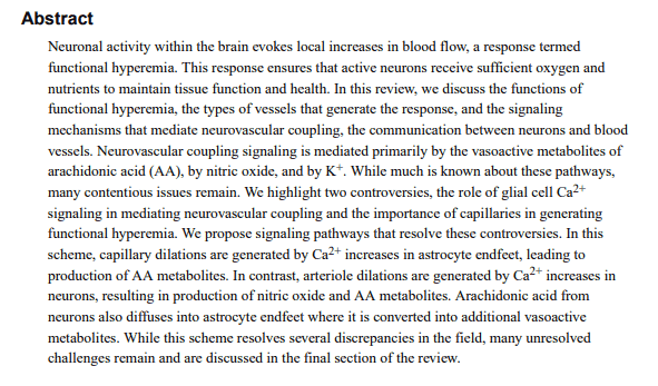
Increased neuronal activity within specific cortical regions elevates local metabolic demand and triggers regional increases in cerebral blood flow, a phenomenon known as functional hyperemia.
Sensory stimulation, motor activity, cognitive processing, and seizure activity all increase regional metabolism and consequently enhance blood flow to activated brain areas.
This neurovascular coupling ensures adequate oxygen and glucose delivery to metabolically active neurons.
Metabolic Vasodilators
Several metabolites have been proposed as mediators of metabolic cerebrovascular regulation, including:
- Adenosine
- Hydrogen ions
- Potassium ions
Adenosine
Adenosine is considered one of the strongest candidates for metabolic flow regulation. Brain adenosine concentration rises rapidly during ischemia, hypoxia, and increased neuronal activity. Adenosine acts as a potent vasodilator on cerebral vessels and contributes significantly to metabolic hyperemia.
Potassium Ions
Enhanced neuronal activity increases extracellular potassium concentration, which promotes vasodilation of pial arterioles.
Hydrogen Ions
Increased metabolic activity and seizure states elevate extracellular hydrogen ion concentration, further contributing to local vasodilation.
Autoregulation of Cerebral Blood Flow
Principles of Cerebral Autoregulation
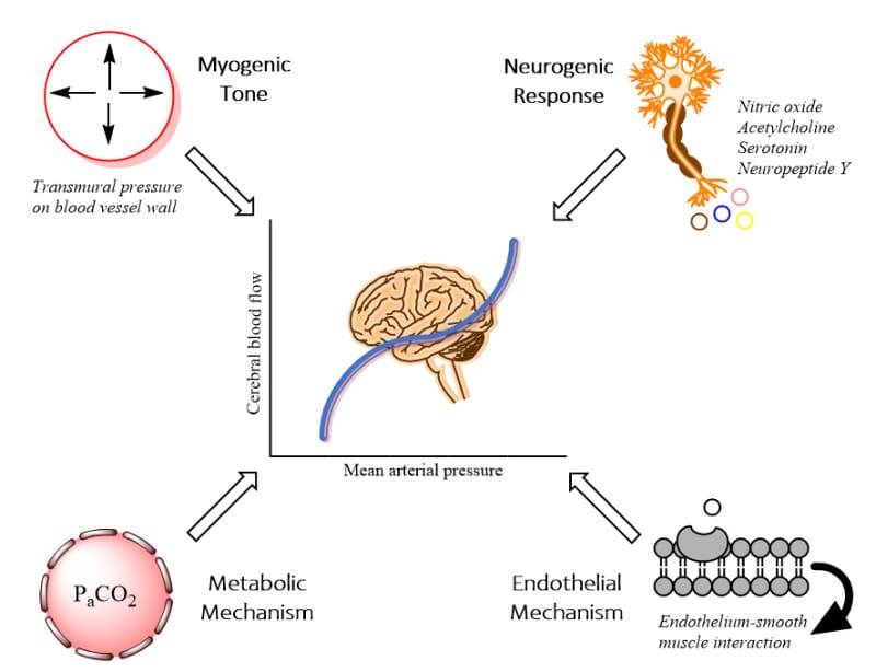
Cerebral autoregulation refers to the intrinsic ability of cerebral vessels to maintain relatively constant blood flow despite fluctuations in arterial blood pressure.
When arterial pressure rises, cerebral precapillary vessels constrict, increasing cerebrovascular resistance and limiting excessive increases in blood flow. Conversely, decreased arterial pressure induces vasodilation and reduced resistance.
This autoregulatory mechanism preserves stable cerebral perfusion across a broad physiological pressure range.
Vascular Responses During Pressure Changes
Autoregulatory responses vary according to vessel size and vascular tone.
- Larger pial arterioles play a major role in autoregulatory constriction during hypertension.
- Smaller arterioles exhibit greater dilation during hypotension.
- Extremely high pressures may overcome active vasoconstriction, leading to passive vascular distension and excessive cerebral blood flow.
- Very low pressures may produce passive vessel collapse and reduced perfusion.
Metabolic Versus Myogenic Mechanisms
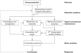
Two principal theories have been proposed to explain cerebral autoregulation:
Myogenic Theory
The myogenic mechanism proposes that vascular smooth muscle responds directly to changes in transmural pressure. Increased pressure induces vasoconstriction, whereas decreased pressure promotes vasodilation.
Metabolic Theory
The metabolic hypothesis suggests that changes in blood flow alter tissue oxygenation and metabolite concentration, which subsequently regulate vascular tone.
Current evidence strongly supports predominant involvement of metabolic mechanisms in cerebral autoregulation.
Experimental studies demonstrate that hypotension-induced vasodilation can be markedly reduced by increasing local oxygen availability, indicating that tissue hypoxia and metabolite accumulation are key mediators of autoregulatory responses.
Adenosine appears to be a major contributor to these metabolic adjustments because its concentration rises rapidly during hypotension and ischemia.
Hydrogen ion concentration does not appear to play a major role in autoregulatory vasodilation because brain pH remains relatively stable during hypotension.
Conclusion
Cerebral circulation is regulated through highly integrated neurogenic, chemical, metabolic, and autoregulatory mechanisms that ensure continuous and stable perfusion of brain tissue. Among these mechanisms, local metabolic and chemical factors exert the strongest influence on cerebral vascular tone, whereas neurogenic control appears to play a more limited modulatory role.
Carbon dioxide, oxygen availability, adenosine, potassium ions, and local metabolic activity are central regulators of cerebrovascular function. In addition, cerebral autoregulation protects neural tissue against ischemia and hypertensive vascular injury by dynamically adjusting vascular resistance in response to pressure fluctuations.
A deeper understanding of these regulatory pathways remains essential for advancing the diagnosis and treatment of neurological disorders such as stroke, traumatic brain injury, cerebral ischemia, hypertension-associated encephalopathy, and neurodegenerative diseases.
