PD-1/PD-L1 Signaling in Disease and Cancer Immunotherapy
Introduction to the PD-1/PD-L1 Immune Checkpoint Pathway
The human immune system is a highly coordinated defense network responsible for protecting the body against pathogens such as bacteria, viruses, fungi, and parasites. In addition to combating infections, the immune system also contributes to the identification and elimination of abnormal or mutated cells. This concept, known as immune surveillance, explains how immune cells recognize and destroy potentially cancerous cells before tumor development occurs.
One of the most important regulatory mechanisms involved in immune control is the programmed death-1 (PD-1) and programmed death-ligand 1 (PD-L1) signaling pathway. This immune checkpoint pathway plays a critical role in maintaining immune tolerance, preventing excessive inflammation, and regulating T-cell activity. However, abnormal activation of the PD-1/PD-L1 axis is strongly associated with multiple diseases, including cancer, autoimmune disorders, inflammatory diseases, and neurological conditions.
Tumor cells exploit this pathway to evade immune destruction by suppressing cytotoxic T-cell responses. Because of this, PD-1 and PD-L1 have become major targets in modern cancer immunotherapy.

Structure and Biological Function of PD-1 and PD-L1
PD-1, also known as CD279, is an inhibitory receptor expressed mainly on activated immune cells including:
- T lymphocytes
- B lymphocytes
- Natural killer (NK) cells
- Natural killer T (NKT) cells
- Myeloid cells
Its ligands include:
- PD-L1 (CD274 or B7-H1)
- PD-L2 (CD273 or B7-DC)
PD-L1 is broadly expressed on both immune and non-immune cells, including:
- Antigen-presenting cells (APCs)
- Dendritic cells
- Macrophages
- Endothelial cells
- Epithelial cells
- Tumor cells
- Activated lymphocytes
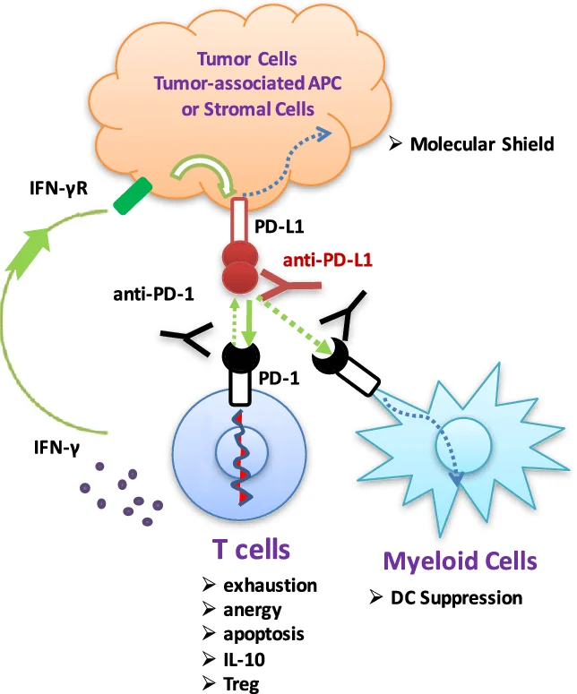
PD-L1 expression is strongly induced by inflammatory cytokines, particularly interferon-gamma (IFN-γ).
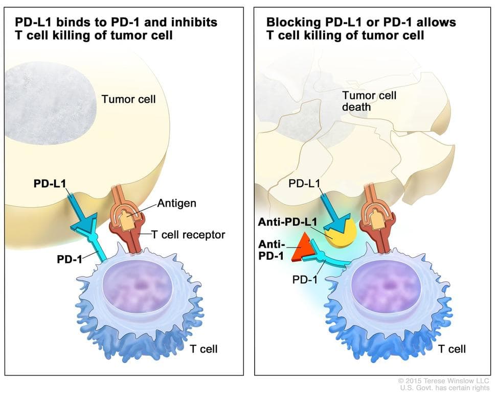
When PD-L1 binds to PD-1 on activated T cells, inhibitory intracellular signaling pathways are activated, leading to:
- Reduced T-cell proliferation
- Decreased cytokine production
- T-cell exhaustion
- Suppression of cytotoxic activity
- Induction of apoptosis in immune cells
This mechanism is physiologically important for preventing autoimmune damage and maintaining peripheral immune tolerance. However, cancer cells hijack this pathway to suppress anti-tumor immunity and survive within the host.
Role of PD-1/PD-L1 in Tumor Immune Evasion
Cancer cells use multiple mechanisms to escape immune surveillance. One of the most effective strategies involves overexpression of PD-L1 on tumor cell surfaces.
Binding of tumor PD-L1 to PD-1 receptors on T cells suppresses immune activation and allows tumor cells to avoid destruction. In addition, many tumors reduce expression of major histocompatibility complex class I (MHC-I) molecules, preventing antigen recognition by cytotoxic T cells.
Other immune escape mechanisms include:
- Secretion of immunosuppressive cytokines such as TGF-β and IL-10
- Production of vascular endothelial growth factor (VEGF)
- Reduction of co-stimulatory molecules
- Recruitment of regulatory T cells (Tregs)
- Activation of myeloid-derived suppressor cells (MDSCs)
These combined mechanisms create an immunosuppressive tumor microenvironment that promotes tumor progression, metastasis, and resistance to therapy.
PD-L1 Expression in Human Cancers
Elevated PD-L1 expression has been identified in many malignancies, including:
- Breast cancer
- Non-small-cell lung carcinoma (NSCLC)
- Melanoma
- Gastric cancer
- Renal cell carcinoma
- Head and neck cancers
- Soft tissue sarcomas
- Endometrial cancer
In several cancers, high PD-L1 expression is associated with:
- Advanced tumor stage
- Increased tumor proliferation
- Reduced survival rates
- Poor clinical prognosis
For example, triple-negative breast cancer frequently exhibits high PD-L1 expression, making it an important target for immune checkpoint blockade therapy.
Interestingly, some studies demonstrate that PD-L1 expression may also correlate with increased tumor-infiltrating lymphocytes (TILs) and improved survival in certain cancer subtypes. This indicates that the prognostic significance of PD-L1 depends on the immune context of the tumor microenvironment.
Molecular Regulation of PD-L1 Expression
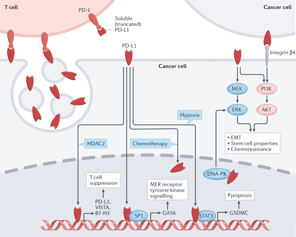
Tumor cells regulate PD-L1 expression through several oncogenic signaling pathways, including:
- JAK/STAT pathway
- PI3K/AKT/mTOR signaling
- MAPK pathway
- NF-κB pathway
- MEK/ERK signaling
Inflammatory cytokines such as IFN-γ and TNF-α also stimulate PD-L1 expression within tumors.
These signaling pathways contribute to immune resistance and enhance tumor survival under immune pressure.
PD-1/PD-L1 and Autoimmune Diseases
The PD-1/PD-L1 pathway is essential for preventing autoimmunity by suppressing excessive immune activation against self-antigens.
Loss or dysfunction of PD-1 signaling can result in severe autoimmune diseases. Experimental studies using PD-1 knockout mice demonstrate spontaneous autoimmune disorders, including:
- Lupus-like nephritis
- Autoimmune arthritis
- Cardiomyopathy
- Type 1 diabetes
Type 1 Diabetes
In non-obese diabetic (NOD) mouse models, blocking PD-1 or PD-L1 accelerates autoimmune destruction of pancreatic beta cells.
Reduced PD-1 expression promotes:
- Increased T-cell infiltration
- Enhanced inflammatory cytokine production
- Severe insulitis
- Earlier onset of diabetes
Human studies also show reduced PD-1 expression on CD4+ T cells in autoimmune diabetic patients.
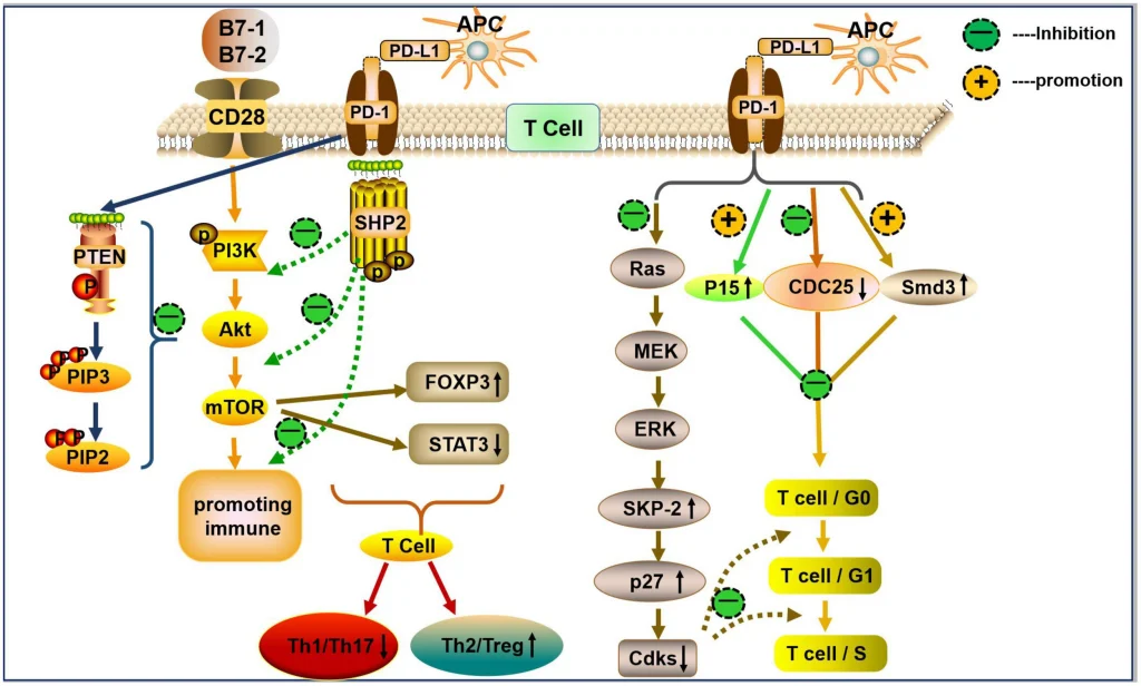
Multiple Sclerosis and PD-1/PD-L1 Signaling
Multiple sclerosis (MS) is a chronic autoimmune disease affecting the central nervous system.
PD-1 and PD-L1 regulate immune responses within the brain and spinal cord by suppressing autoreactive T cells.
In experimental autoimmune encephalomyelitis (EAE), an animal model of MS:
- PD-1 deficiency worsens neurological inflammation
- Increased levels of IL-17, TNF-α, IFN-γ, and IL-6 are observed
- Disease progression accelerates
PD-L1 expression is elevated in MS lesions, suggesting an attempt by the body to suppress excessive immune-mediated damage.
Inflammatory Bowel Disease and Intestinal Immunity
The PD-1/PD-L1 pathway also contributes to intestinal immune tolerance.
In inflammatory bowel disease (IBD), including Crohn’s disease and ulcerative colitis:
- PD-1 expression increases on intestinal T cells
- PD-L1 is elevated in inflamed colon tissues
- Disruption of PD-1 signaling enhances intestinal inflammation
Loss of PD-L1 activity leads to:
- Increased Th1 inflammatory responses
- Epithelial barrier disruption
- Overgrowth of intestinal bacteria
- Severe tissue injury
These findings demonstrate the importance of PD-1/PD-L1 in maintaining gastrointestinal immune homeostasis.
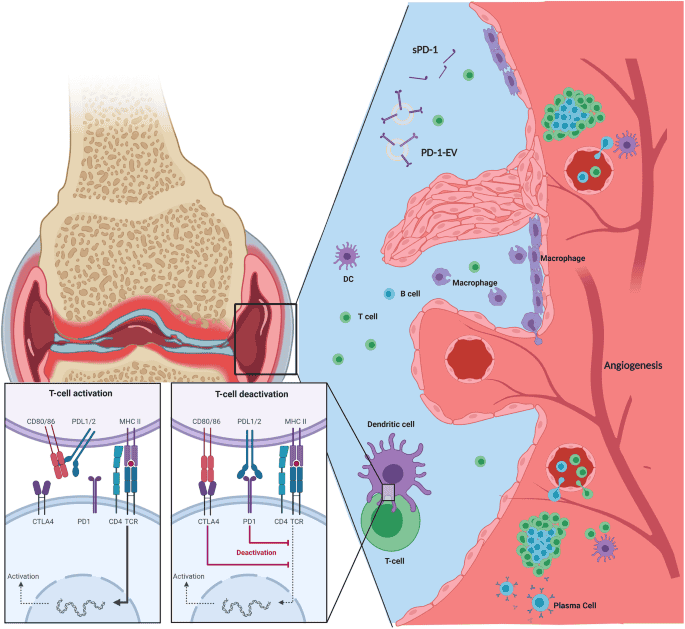
PD-1/PD-L1 in Rheumatoid Arthritis
Rheumatoid arthritis (RA) is characterized by chronic joint inflammation and immune dysregulation.
Patients with RA frequently exhibit:
- Elevated soluble PD-1 levels
- Increased PD-1 expression on synovial T cells
- Enhanced PD-L1 expression on dendritic cells and macrophages
Experimental studies indicate that PD-1 deficiency exacerbates arthritis severity through increased inflammatory cytokine secretion and T-cell activation.
Neurological Disorders and Neuroinflammation
PD-1/PD-L1 signaling participates in regulating neuroinflammatory responses in conditions such as:
- Alzheimer’s disease
- Stroke
- Multiple sclerosis
In Alzheimer’s disease:
- Reduced PD-1 expression on T cells is observed
- Decreased PD-L1 expression on macrophages correlates with lower IL-10 production
Because IL-10 has neuroprotective anti-inflammatory functions, impaired PD-1/PD-L1 signaling may contribute to neurodegeneration.
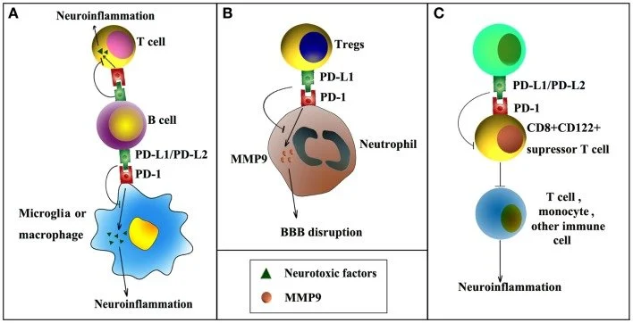
PD-1/PD-L1 in Stroke
Following ischemic stroke, immune regulation becomes critical for limiting secondary brain injury.
PD-L1 expression on B cells suppresses inflammatory activation of:
- T cells
- Microglia
- Macrophages
- Neutrophils
Experimental models show that PD-L1 deficiency worsens post-stroke inflammation and neurological damage.
This suggests that PD-1/PD-L1 signaling may serve neuroprotective functions during cerebral ischemia.
Immune Checkpoint Inhibitors in Cancer Therapy
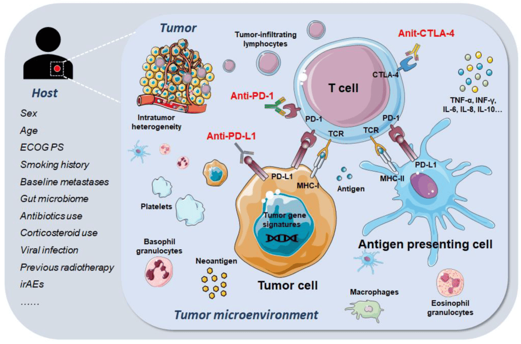
The discovery of immune checkpoint pathways revolutionized cancer treatment. Blocking PD-1 or PD-L1 restores T-cell activity against tumors and enhances anti-cancer immunity.
Major FDA-approved immune checkpoint inhibitors include:
PD-1 Inhibitors
- Nivolumab
- Pembrolizumab
PD-L1 Inhibitors
- Atezolizumab
These therapies are widely used in:
- Melanoma
- NSCLC
- Renal cell carcinoma
- Gastric cancer
- Urothelial carcinoma
- Triple-negative breast cancer
Immune checkpoint inhibitors significantly improve survival in many advanced cancers.
Adverse Effects of PD-1/PD-L1 Blockade
Although checkpoint inhibitors are highly effective, they can also trigger immune-related adverse events due to excessive immune activation.
Reported toxicities include:
- Colitis
- Pneumonitis
- Thyroid dysfunction
- Nephritis
- Autoimmune diabetes
- Neurological complications
These side effects are generally manageable with immunosuppressive therapies such as corticosteroids.
PD-L1 as a Biomarker in Precision Oncology
Accurate measurement of PD-L1 expression is essential for selecting patients likely to respond to checkpoint blockade therapy.
Current approaches include:
- Immunohistochemistry (IHC)
- Radiolabeled anti-PD-L1 imaging
- Molecular profiling
However, PD-L1 expression is highly heterogeneous within tumors, making standardized assessment challenging.
Future precision oncology approaches may combine:
- PD-L1 status
- Tumor mutational burden
- Immune cell infiltration profiles
- Genomic biomarkers
to improve therapeutic decision-making.
Future Perspectives of PD-1/PD-L1 Research
Research into PD-1/PD-L1 biology continues to expand rapidly.
Future developments may include:
- Combination immunotherapies
- Personalized cancer vaccines
- Enhanced immune checkpoint modulation
- Gene-editing approaches
- Biomarker-guided therapies
- Novel inhibitors targeting multiple immune suppressive pathways
Understanding how PD-1/PD-L1 signaling interacts with the tumor microenvironment, microbiome, metabolism, and host genetics will improve next-generation immunotherapies for cancer, autoimmune diseases, and neurological disorders.
Conclusion
The PD-1/PD-L1 immune checkpoint pathway is a central regulator of immune tolerance and immune suppression. Under normal physiological conditions, this signaling system protects tissues from excessive inflammation and autoimmunity. However, many diseases exploit or disrupt this pathway.
In cancer, overexpression of PD-L1 enables tumor cells to escape immune destruction and promotes tumor progression. Conversely, reduced PD-1 signaling contributes to autoimmune and inflammatory diseases.
The development of immune checkpoint inhibitors targeting PD-1 and PD-L1 has transformed cancer therapy and established immunotherapy as a major pillar of modern medicine. Continued research into immune checkpoint biology will provide new opportunities for precision medicine, improved immunotherapeutics, and personalized treatment strategies across multiple disease states.
