Purification, Characterization, and Biological Role of Tumor-Derived Exosomes
Introduction to Tumor-Derived Exosomes
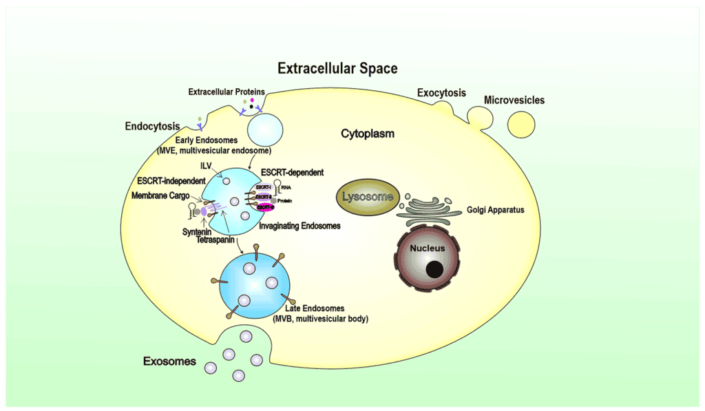
Exosomes are nanosized extracellular vesicles released by many cell types, including cancer cells. These membrane-bound vesicles originate from the endosomal pathway and typically range from 30 to 100 nanometers in diameter. Under electron microscopy, exosomes display a characteristic cup-shaped or saucer-like morphology surrounded by a lipid bilayer membrane. Because of their small size and molecular composition, exosomes are now recognized as important mediators of intercellular communication in both normal physiology and cancer biology.
Tumor-derived exosomes (Tu-exosomes) contain a wide variety of biologically active molecules, including membrane proteins, heat-shock proteins, major histocompatibility complex (MHC) molecules, oncogenic receptors, lipids, enzymes, and nucleic acids. Previous studies have demonstrated that exosomes secreted by dendritic cells, commonly called dexosomes, can modulate immune responses through interactions between MHC molecules and T-cell receptors. In cancer research, tumor-secreted exosomes have attracted major interest because they contribute to tumor progression, metastasis, angiogenesis, immune modulation, and cellular signaling.
Recent investigations have shown that exosomes released from tumor cells may contain tumor-associated proteins such as HER2/neu, a receptor frequently overexpressed in aggressive breast cancers. The detection of tumor-specific markers on exosomes suggests that these vesicles may serve as biomarkers for cancer diagnosis and monitoring, as well as potential therapeutic targets.
Biological Importance of Tumor-Derived Exosomes
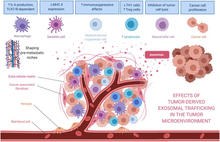
Tumor-derived exosomes play multiple roles in the tumor microenvironment and systemic cancer progression. These extracellular vesicles are involved in:
- Cell-to-cell communication
- Transfer of oncogenic proteins
- Regulation of immune responses
- Promotion of angiogenesis
- Facilitation of tumor invasion and metastasis
- Modification of surrounding stromal tissues
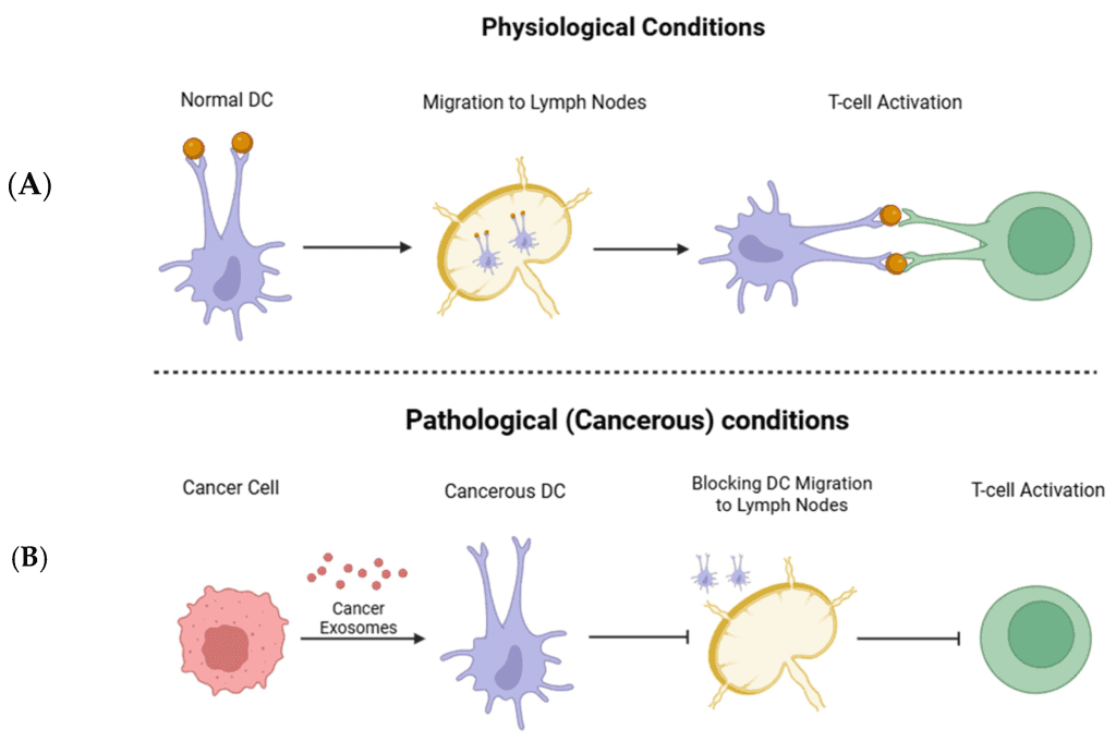
Studies have demonstrated that exosomes derived from platelets and tumor cells can stimulate angiogenesis and metastatic dissemination, especially in lung and breast cancers. In addition, tumor exosomes can transport antigens from malignant cells to antigen-presenting cells such as dendritic cells, potentially influencing antitumor immune responses.
Because tumor exosomes contain membrane proteins and signaling molecules originating from the parental cancer cell, they are considered valuable tools for studying tumor biology and developing cancer immunotherapies.
Challenges in Exosome Isolation and Purification
Accurate investigation of tumor-derived exosomes requires highly purified exosome preparations. Conventional exosome isolation methods generally involve sequential centrifugation and ultracentrifugation steps designed to eliminate cells, cellular debris, and larger extracellular vesicles.
The standard purification process includes:
- Low-speed centrifugation to remove cells
- Intermediate centrifugation to eliminate debris
- Filtration through microporous membranes
- High-speed ultracentrifugation to pellet exosomes
Although these approaches successfully enrich exosome populations, they often produce preparations with limited purity because contaminants such as protein aggregates and microvesicles remain present. Previous protocols reported exosome purities below 70%, limiting functional and molecular analyses.
To improve specificity and purity, researchers developed immunoaffinity-based purification methods targeting tumor-associated proteins expressed on exosome membranes.
HER2-Targeted Isolation of Tumor Exosomes
A novel purification strategy was developed using HER2-targeted immunomagnetic beads to selectively isolate HER2-positive tumor exosomes. HER2 is a transmembrane receptor overexpressed in several aggressive cancers, particularly HER2-positive breast carcinoma.
The method utilized Herceptin (Trastuzumab), a monoclonal antibody against HER2, coupled to protein G-coated paramagnetic beads. These antibody-coated beads specifically bind HER2-expressing exosomes, enabling highly selective isolation.
Two human breast cancer cell lines were used:
- BT-474 cells with strong HER2 overexpression
- MDA-MB-231 cells with low HER2 expression
The experimental results demonstrated that HER2-positive exosomes strongly attached to Herceptin-coated beads, while low HER2-expressing cells showed minimal binding activity. Flow cytometry analysis confirmed nearly complete binding efficiency of HER2-containing exosomes.
This immunomagnetic separation approach significantly improved exosome purification specificity compared with conventional ultracentrifugation alone.
Exosome Isolation Procedure
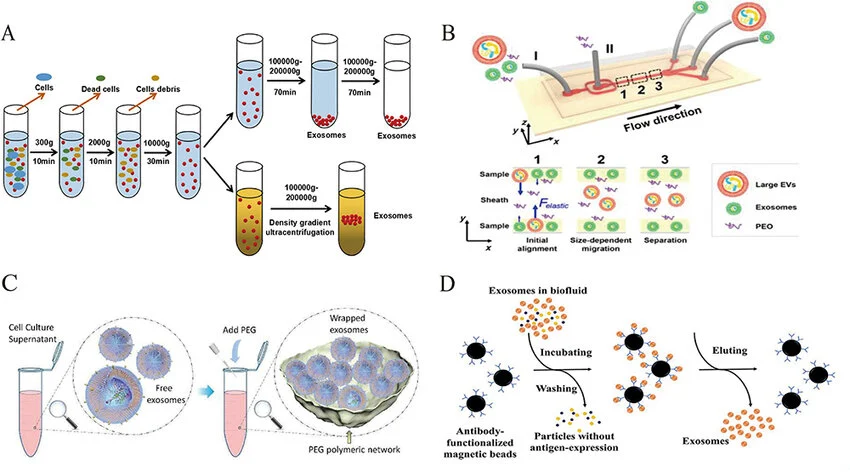
The purification workflow consisted of several critical laboratory steps:
Cell Culture Preparation
Tumor cells were maintained in RPMI 1640 culture medium supplemented with fetal bovine serum previously depleted of bovine exosomes through ultracentrifugation.
Sequential Centrifugation
Cell culture supernatants underwent multiple centrifugation stages:
- 300 × g to remove intact cells
- 1,200 × g to remove debris
- Filtration through 0.2 µm filters
- Ultracentrifugation at 100,000 × g to pellet exosomes
Density Cushion Purification
The exosome pellet was purified further using a sucrose/deuterium oxide density cushion, improving vesicle enrichment and removing contaminants.
Immunomagnetic Separation
Purified exosomes were incubated with Herceptin-coated magnetic beads overnight. Magnetic separation allowed selective recovery of HER2-positive tumor exosomes.
Characterization of Tumor-Derived Exosomes
Several analytical methods were used to characterize isolated exosomes.
Flow Cytometry (FACS)
Fluorescence-activated cell sorting confirmed the presence of HER2 protein on isolated exosomes. HER2-positive exosomes displayed strong and uniform fluorescence intensity after labeling with FITC-conjugated anti-HER2 antibodies.
Western Blot Analysis
Western blotting verified the expression of HER2 protein in exosome preparations. Protein lysates from cells and exosomes were separated using SDS-PAGE and transferred onto nitrocellulose membranes for antibody-based detection.
Electron Microscopy
Transmission electron microscopy revealed classical exosome morphology, including:
- Cup-shaped membrane vesicles
- Lipid bilayer structure
- Diameter ranging from 30 to 120 nm
These findings confirmed successful purification of authentic tumor-derived exosomes.
Isolation of Exosomes from Malignant Ascites
Researchers also applied the HER2-targeted purification strategy to malignant ascitic fluid obtained from a patient with advanced ovarian cancer.
Exosomes isolated from ascites contained HER2-positive vesicles detectable by flow cytometry. Herceptin-coated beads successfully captured these tumor-associated exosomes, demonstrating the potential clinical utility of antibody-based exosome isolation directly from patient-derived biological fluids.
This approach may facilitate:
- Cancer biomarker discovery
- Liquid biopsy development
- Personalized cancer diagnostics
- Exosome-based immunotherapy research
Functional Effects of Tumor Exosomes on Cancer Cells
To investigate whether tumor-derived exosomes possess biological activity, researchers studied their effects on BT-474 breast cancer cell proliferation.
Fluorescence microscopy demonstrated that labeled exosomes attached directly to the surface of HER2-positive cancer cells. Functional assays using MTT analysis showed that tumor-derived exosomes significantly increased cancer cell proliferation.
These observations suggest that exosomes may stimulate signaling pathways involved in:
- Cell cycle progression
- Tumor growth
- Cellular communication
- Oncogenic signaling
The ability of exosomes to transfer membrane proteins, ligands, and signaling molecules between cells may contribute to cancer progression and resistance mechanisms.
Potential Mechanisms of Exosome-Mediated Tumor Progression
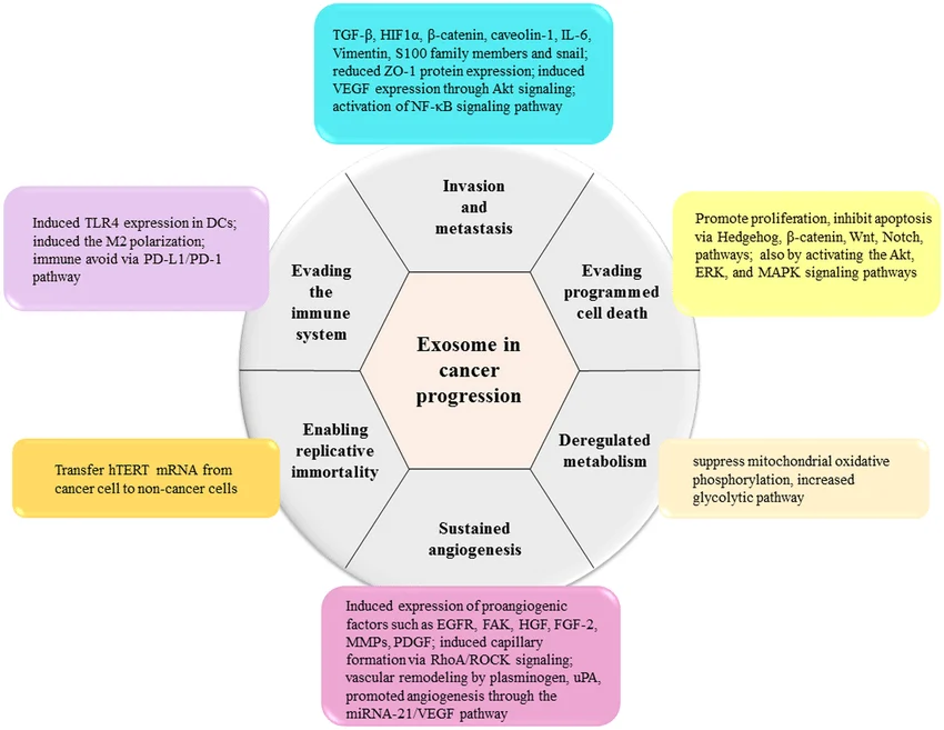
Tumor exosomes may influence recipient cells through several mechanisms:
Membrane Fusion
Exosomes can fuse with target cell membranes and transfer proteins, receptors, and lipids directly into recipient cells.
Receptor-Ligand Signaling
Surface molecules on exosomes may activate cellular receptors and downstream signaling cascades involved in proliferation and survival.
Transfer of Genetic Material
Exosomes can deliver mRNA, microRNA, and other nucleic acids that alter gene expression in recipient cells.
Immune System Modulation
Tumor-derived exosomes may suppress antitumor immunity or modulate dendritic cell activity.
These mechanisms contribute to the development of an aggressive tumor microenvironment favorable to cancer progression.
Clinical and Therapeutic Applications of Exosome Research
The selective purification of tumor-derived exosomes opens new opportunities in oncology and precision medicine.
Potential applications include:
- Early cancer detection
- Biomarker discovery
- Monitoring therapeutic response
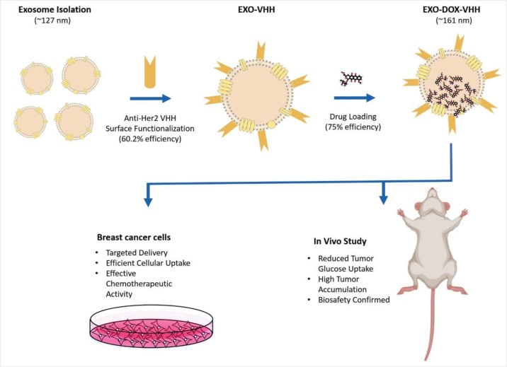
- Drug delivery systems
- Cancer vaccine development
- Personalized immunotherapy
Because tumor exosomes contain tumor-associated antigens, they may serve as valuable antigen sources for dendritic cell-based vaccines and immune activation strategies.
Antibody-coated magnetic bead systems targeting multiple tumor antigens could improve selective isolation of clinically relevant exosome populations from blood, ascites, pleural effusions, and other biological samples.
Conclusion
Tumor-derived exosomes are biologically active extracellular vesicles that play essential roles in cancer progression, intercellular communication, immune regulation, and metastasis. Their molecular composition reflects the characteristics of the parental tumor cells, making them promising biomarkers and therapeutic targets.
The development of HER2-targeted immunomagnetic purification methods represents an important advancement in exosome research. This technique enables highly selective isolation of tumor-associated exosomes from both cultured cells and patient-derived fluids.
Experimental findings indicate that tumor-derived exosomes can directly stimulate cancer cell proliferation, supporting their role in tumor growth and progression. Continued investigation into exosome biology may lead to innovative diagnostic tools and novel therapeutic strategies for cancer treatment.
